


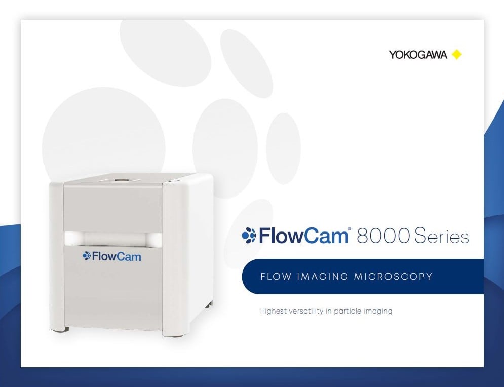
Flow Imaging Microscopy
Characterize Particles & Organisms with Confidence
Obtain the highest-resolution images in real time. Analyze the shape, size, and count of subvisible particles, cells, and plankton with flow imaging microscopy.
FlowCam. Particle Analysis with Vision®.
FlowCam Applications
For Sample Analysis That Demands Detail
Leading flow imaging analysis for applications where morphological characterization with high-quality images is a must – including oceanographic research, municipal water quality monitoring, biophysical analysis of API aggregates, cell and gene therapy, organoids, food and beverage analysis, materials characterization, and many other areas of research.
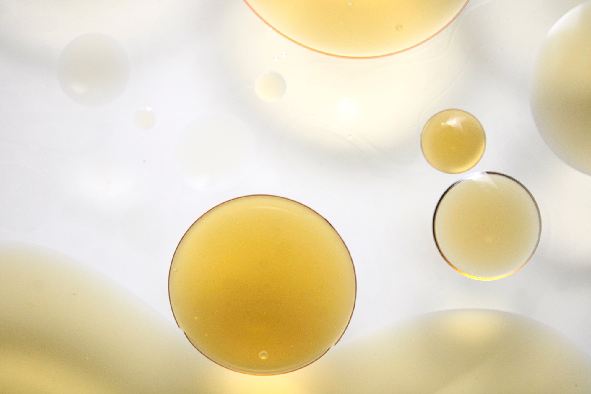
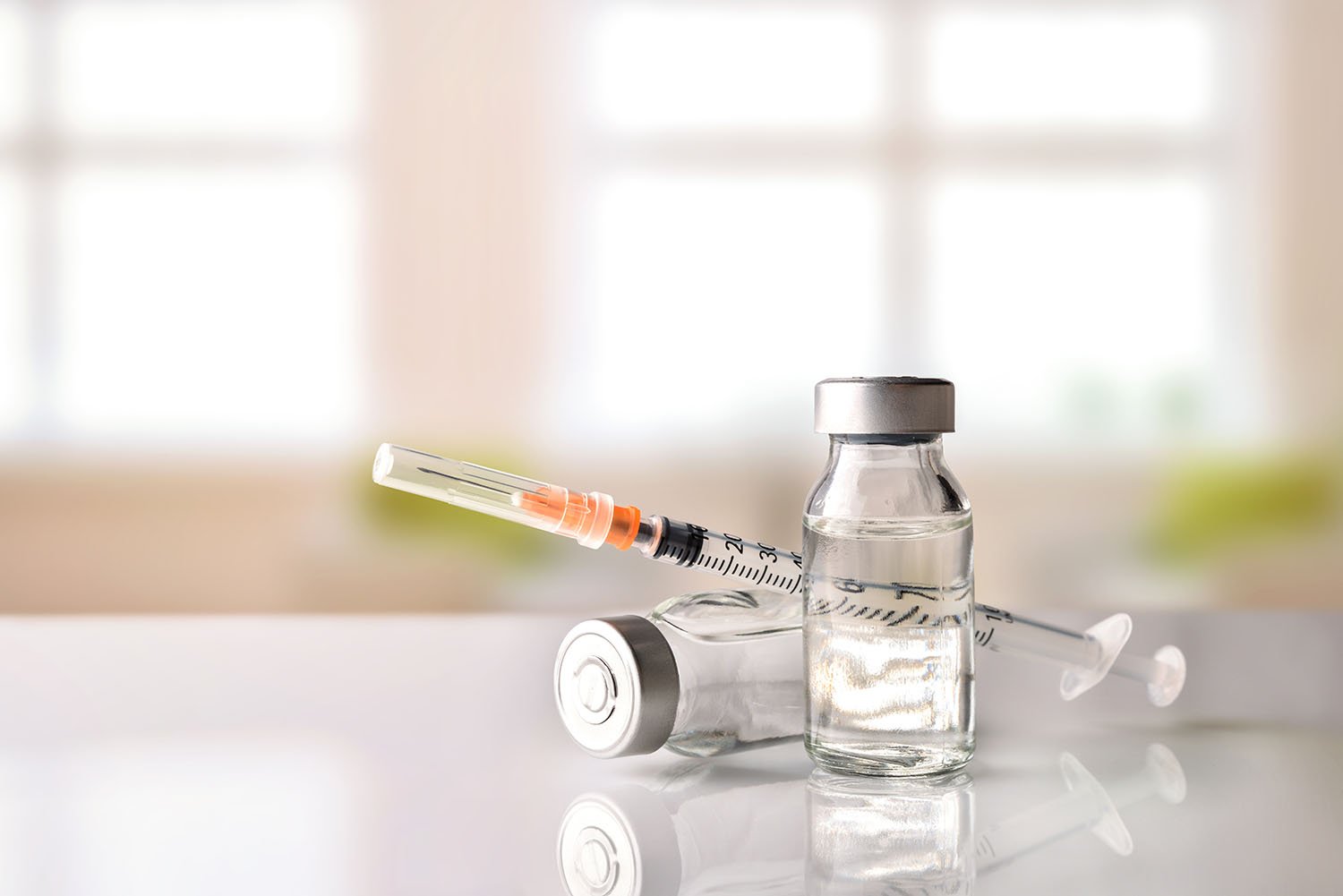
Ensure the safety and efficacy of your biologic drug products
For the most reliable results and a complete picture of safety and efficacy, USP <1788> recommends flow imaging microscopy as an orthogonal method to Light Obscuration for determining subvisible particulate content in parenteral drug formulations.
With its ability to produce higher-quality images than any other flow imaging microscopy instrument in the industry, FlowCam has become an indispensable tool in biotherapeutics research, formulation, and development for proteins, nano-drug delivery systems, organoids, and cell and gene therapy products.

Learn more about how flow imaging microscopy with FlowCam can characterize subvisible and submicron API aggregates and other particulates using morphology and artificial intelligence.
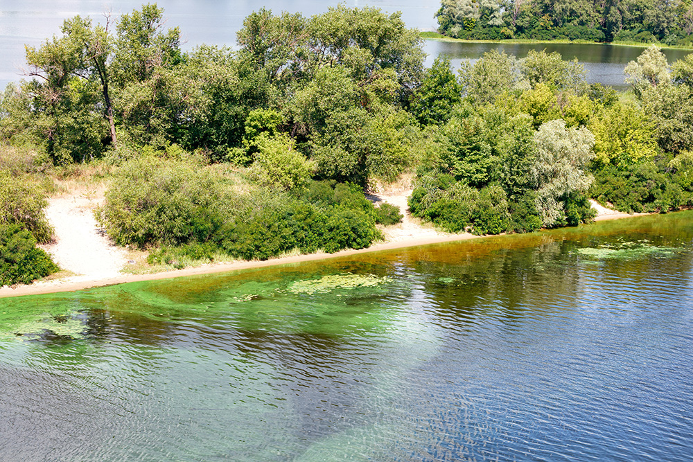
Add speed and reliability to your microplankton analysis
FlowCam is the fast and accurate alternative to manual microscopy for quantifying phytoplankton and zooplankton biovolume and community composition, monitoring source water, and identifying cyanobacteria and harmful algal blooms.
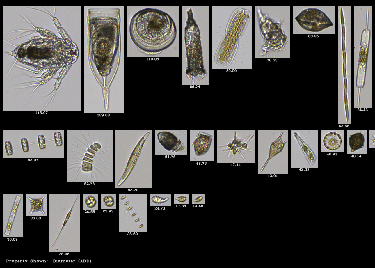
Flow imaging microscopy combines high-resolution digital imaging with precise fluidics for rapid, automated analysis of water samples. It provides statistical data on plankton and particle morphology, helping researchers identify, classify, and quantify diverse microorganism populations with exceptional detail and reproducibility.

Discover how FlowCam is used in marine and freshwater applications, aquaculture, and microalgae cultivation
Monitor particle shape to improve product quality and performance
Flow imaging microscopy combines particle counting and sizing with morphology to characterize microscopic particles in a wide variety of materials, environmental, and chemical applications – from food and beverage ingredients to crop science, printer toner, fiber analysis, additive manufacturing, and polymer composition analysis.
FlowCam streamlines quality control processes through comprehensive particle characterization. Capturing detailed images of each particle suspended in a liquid, flow imaging microscopy reveals critical morphological attributes that affect product performance and stability in ways laser diffraction and light obscuration methods cannot match.

Learn how Flow Imaging Microscopy works to characterize microparticles in a fluid medium
-
Ensure the safety and efficacy of your biologic drug products
For the most reliable results and a complete picture of safety and efficacy, USP <1788> recommends flow imaging microscopy as an orthogonal method to Light Obscuration for determining subvisible particulate content in parenteral drug formulations.
With its ability to produce higher-quality images than any other flow imaging microscopy instrument in the industry, FlowCam has become an indispensable tool in biotherapeutics research, formulation, and development for proteins, nano-drug delivery systems, organoids, and cell and gene therapy products.
Learn more about how flow imaging microscopy with FlowCam can characterize subvisible and submicron API aggregates and other particulates using morphology and artificial intelligence.
-
Add speed and reliability to your microplankton analysis
FlowCam is the fast and accurate alternative to manual microscopy for quantifying phytoplankton and zooplankton biovolume and community composition, monitoring source water, and identifying cyanobacteria and harmful algal blooms.
Flow imaging microscopy combines high-resolution digital imaging with precise fluidics for rapid, automated analysis of water samples. It provides statistical data on plankton and particle morphology, helping researchers identify, classify, and quantify diverse microorganism populations with exceptional detail and reproducibility.
Discover how FlowCam is used in marine and freshwater applications, aquaculture, and microalgae cultivation
-
Monitor particle shape to improve product quality and performance
Flow imaging microscopy combines particle counting and sizing with morphology to characterize microscopic particles in a wide variety of materials, environmental, and chemical applications – from food and beverage ingredients to crop science, printer toner, fiber analysis, additive manufacturing, and polymer composition analysis.
FlowCam streamlines quality control processes through comprehensive particle characterization. Capturing detailed images of each particle suspended in a liquid, flow imaging microscopy reveals critical morphological attributes that affect product performance and stability in ways laser diffraction and light obscuration methods cannot match.
Learn how Flow Imaging Microscopy works to characterize microparticles in a fluid medium
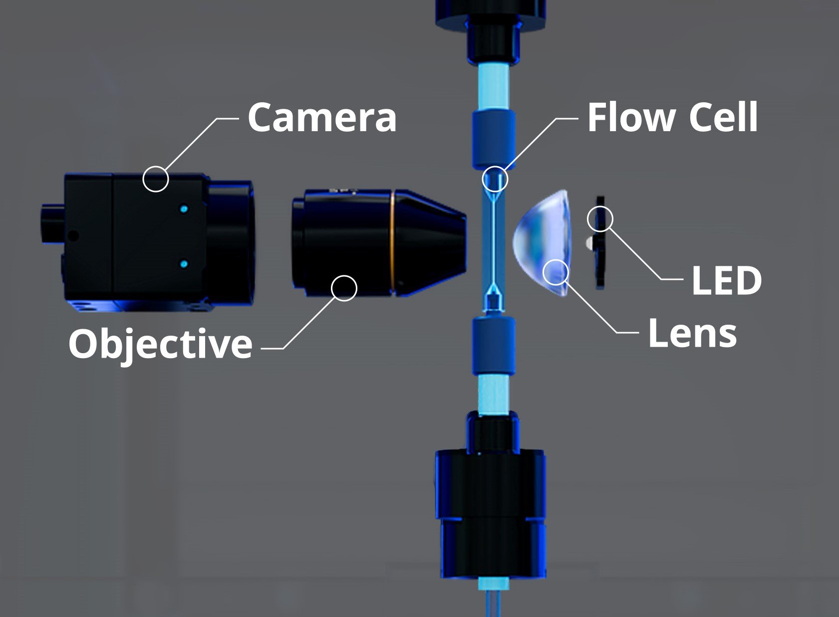
In flow imaging microscopy, particles move through a flow cell as images are captured in real-time by a high-speed camera and processed by image analysis software.
How FlowCam Works
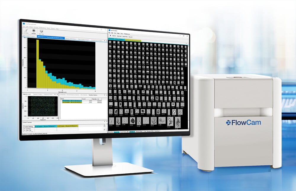
Analyze Particle Count, Size, and Morphology with Flow Imaging Microscopy
FlowCam rapidly acquires digital images of particles to measure particle size and morphology. Up to 50,000 particles/minute may be imaged in real time and analyzed based on more than 40 morphological parameters. Optional artificial intelligence tools for biopharmaceutical applications allow automated particle identification.
Helpful Resources

Recent Articles


See the Power of Particle Analysis with Vision®
-
Get in Touch
Tell us about your application and particle characterization needs.
-
Have a conversation
We're happy to set up a call to discuss your application and answer your questions.
-
Discuss next steps
Expand your knowledge with a seminar, demonstration, sample analysis, or obtain a quote.
