Automate Detection of Cyanobacteria with FlowCam Cyano
How it Works
Fluorescence-based flow imaging microscopy with FlowCam Cyano uses laser excitation to image and identify Cyanobacteria and differentiate them from algae and other particles in aquatic samples. FlowCam Cyano's optical configuration uses a red laser (633 nm) to detect the unique fluorescence emissions from aquatic organisms.
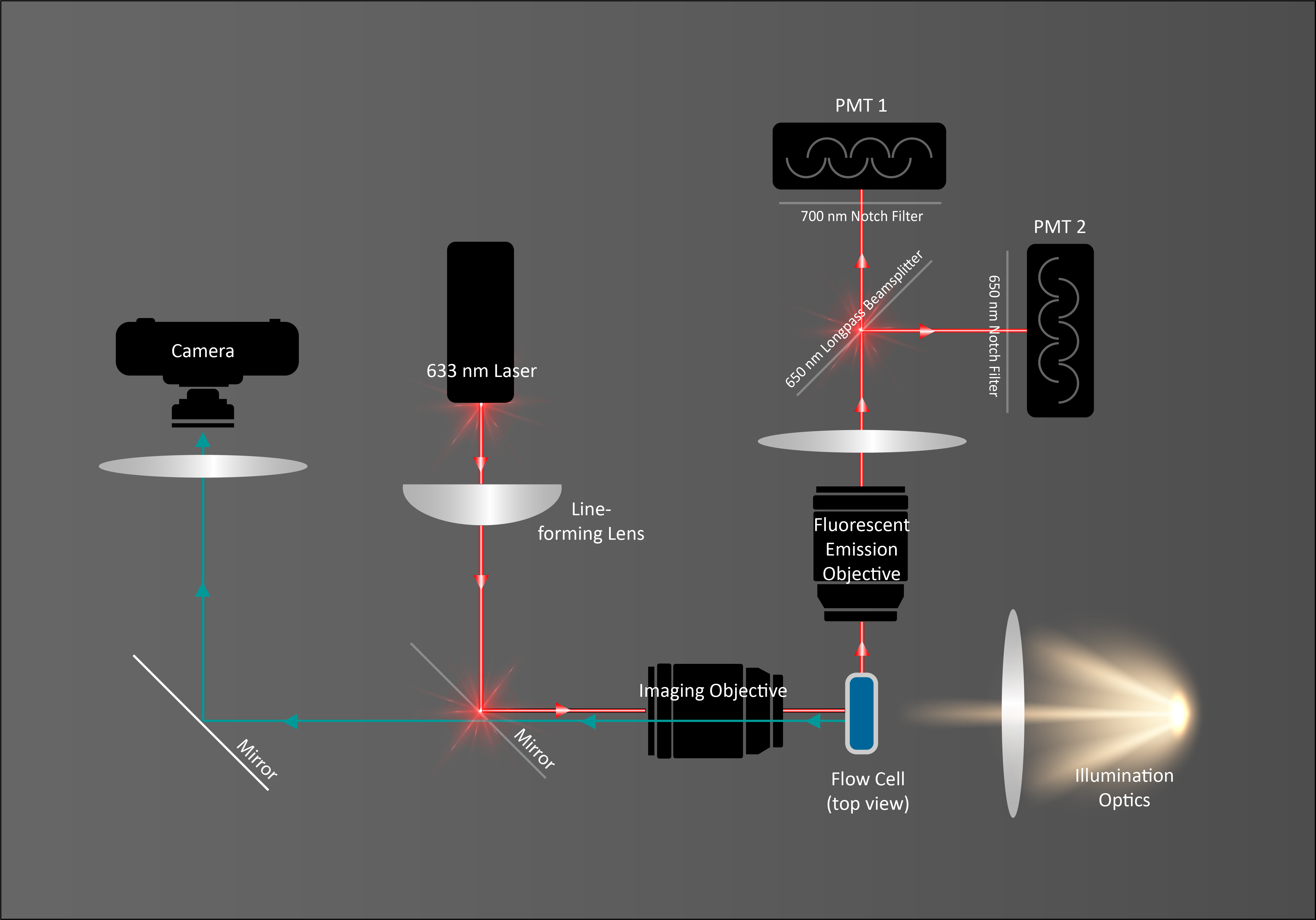
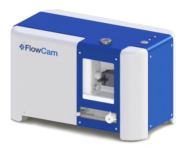
- A sample is manually loaded into the injection port where a high-precision syringe draws the sample into an optical flow cell and a fluidics sensor initiates data acquisition.
- The red laser excites microalgae fluorescence via Channel 1 (chlorophyll) or Channel 2 (phycocyanin) as a sample passes through the flow cell, triggering the camera to capture an image of the entire flow cell.
- FlowCam's image analysis software, VisualSpreadsheet®, isolates each particle or organism as a separate image, and calculates data for each image, including fluorescence ratios, as well as sample count and concentration.
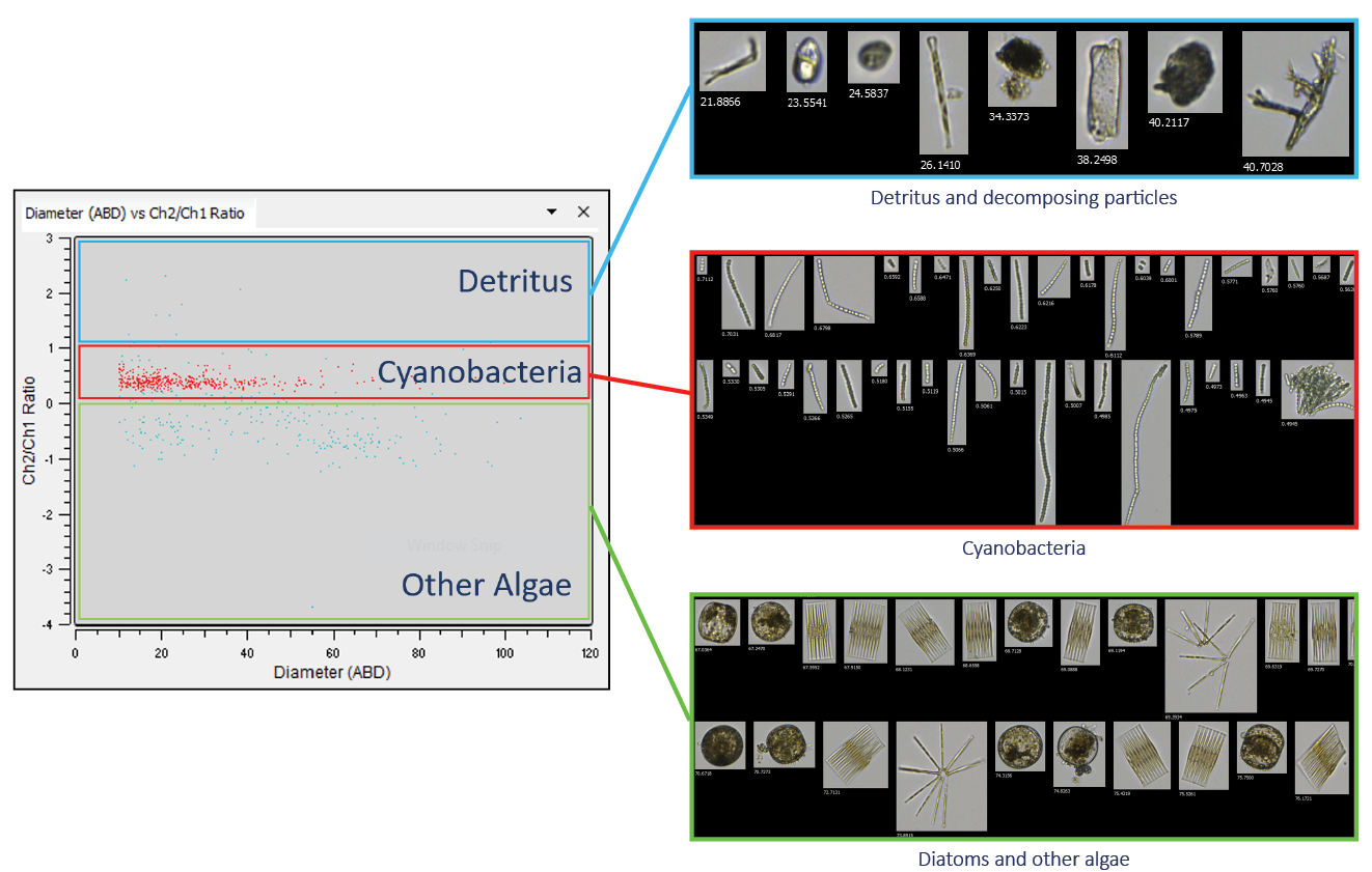
- Pre-built filters sort images into three categories: Cyanobacteria, Diatoms and Other Algae, and Detritus and Decomposing Particles. Data may be further analyzed, grouped, and filtered by the user.
FlowCam Cyano offers three pre-built fluorescent filters to differentiate Cyanobacteria from diatoms and other algae, and detritus and decomposing particles.
Create libraries from microalgae specific to your region to further sort and assist in classification of your count and concentration of different algal populations.

Interested in learning more?
-
Get in Touch
Tell us about your application and particle characterization needs.
-
Have a conversation
We're happy to set up a call to discuss your application and answer your questions.
-
Discuss next steps
Expand your knowledge with a seminar, demonstration, sample analysis, or obtain a quote.


