The FlowCam was recently used by a team led by Natasha Barteneva of Nazarbayev University to develop a method for the classification of Microcystis colonial morphospecies in samples obtained from a long-term mesocosm experiment (the AQUACOSM Lake Mesocosm Warming Experiment).
Microcystis is a freshwater cyanobacteria that occurs worldwide, with detrimental effects that include polution of drinking water supply and harmful impact on plankton, fish, and mollusks. Methods have previously been established for analyzing colonious organisms like Microcystis on the FlowCam.
The team tested a machine learning approach to see if they could bring species-level identification capability to the FlowCam, a flow imaging microscope traditionally used to identify organisms to the genus level.
"We used a benchtop FlowCam imaging cytometer equipped with VisualSpreadsheet software (Yokagawa Fluid Imaging, USA). Samples were recorded in autoimage mode using combinations of 10 × objective (NA = 0.3; resolution 1 pixel equals to 0.554 μm)/100 μm flow cell and/or 20 × objective/50 μm flow cell for identification, classification, and quantification."1
169 mesocosm samples were analyzed, 69 of which contained Microcystis species. 119,135 images of Microcystis were captured by the FlowCam, and 25-50 images of each species were selected from VisualSpreadsheet to be used as representative images. The experimentors also used VisualSpreadsheet's filtering capabilities to determine 48 morphological parameters for both inter- and intra-generic classification. To improve the accuracy needed to differentiate the 5 Microcystis species, the team applied various machine learning algorithms that ranked these image parameters to determine which should be used to best characterize the various species in the development of species-specific filter sets.
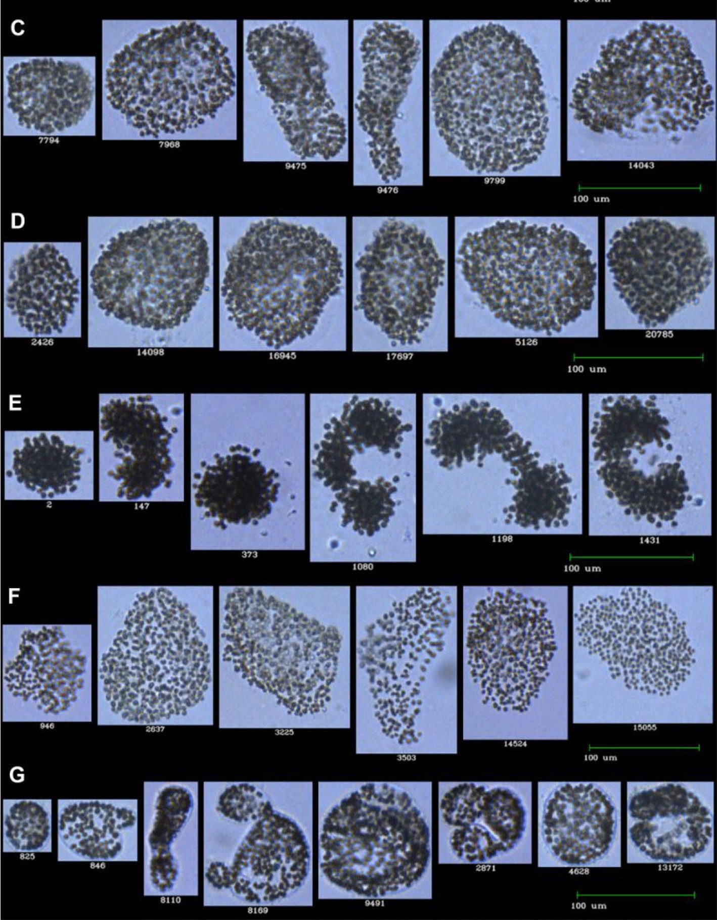
Pictured above: FlowCam images of the 5 Microcystis species observed for the study: (C) Microcystis aeruginosa; (D) M. ichthyoblabe; (E) M. novacekii; (F) M. smithii; (G) M. wesenbergii.
Applying the machine learning filter sets, the FlowCam images were very accurately (96-100%) used to differentiate Microcystis from other genera, and showed some success (67-78%) differentiating between different species of Microcystis (this success rate is approximately equivalent to the success rate of a human taxonomist). This is the first study to differentiate between Microcystis species, and the limited ability to do so by either the FlowCam or a trained scientist is attributed to the high level of Microcystis spp. heterogeneity during seasonal blooms.
"This study demonstrated the proof-of-concept of using a machine learning approach in the analysis of colonial morphospecies of Microcystis. The results described here are based on previous researchers’ work demonstrating the application of machine learning in the identification and counting of different taxa of plankton organisms. The current gold standard for phytoplankton taxonomy is light microscopy of algal samples; however, there is a huge interest to apply semi-automated and automated approaches for Microcystis colonial forms classification. Light microscopy’s biggest disadvantages are the extensive training and time period required for a taxonomist to become a proficient expert, the high cost of training, and the large component of manual work involved. Although sequencing and the following molecular biological identification have become more popular in recent years, microscopy and visual morphological analysis remain the most important and widely available tools. In the context of saving time during taxonomic analysis, imaging cytometers constitute a faster and efficient way to receive the morphological information required for taxonomic identification."1

1Y. Mirasbekov, A. Zhumakhanova, A. Zhantuyakova, K. Sarkytbayev, D.V. Malashenkov, A. Baishulakova, V. Dashkova, T.A. Davisdon, I.A. Vorobjev, E. Jeppesen, N.S. Barteneva. “Semi-Automated classification of colonial Microcystis by FlowCAM imaging flow cytometry in mesocosm experiment reveals high heterogeneity during seasonal bloom” Scientific Reports, May 2021, https://doi.org/10.1038/s41598-021-88661-2.
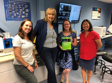
Pictured here: Professor Natasha Barteneva visits the Fluid Imaging lab in 2019 (second from left).


