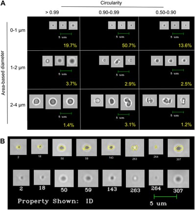
A class of engineered proteins called elastin-like polymers (ELP) have shown promise for advanced  drug delivery applications. In order to fully realize their potential however, they need to be rigorously characterized to determine how they behave in different environments. The FlowCam is ideally suited to help with the characterization process.
Researchers at University of New England (UNE) are the first to use flow imaging microscopy to study ELPs. The FlowCam allowed them to gather tens of thousands of images of individual particles in solution under different conditions. VisualSpreadsheet software provided the ability to sort and filter the images based on different particle characteristics.
Flow Imaging Microscopy has proven to be a useful tool in the analysis of the effects of various solvent conditions on ELP coacervate size, morphology, and behavior, such as the dispersion of single particles versus aggregates.
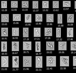
Pictured at right: Images of ELPs as imaged by the FlowCam Nano
The students at UNE worked in conjunction with Nicole Gill, Laboratory Supervisor at Fluid Imaging Technologies. A research paper based on their work was published in PLOS One, a peer-reviewed journal published by the Public Library of Science.
Download the paper here. Flow imaging microscopy as a novel tool for high-throughput evaluation of elastin-like polymer coacervates.


