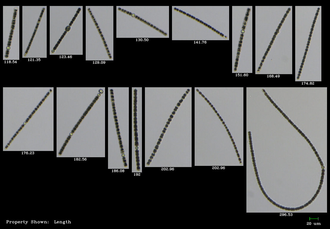
Scientists at the University of Alberta, Alberta Health, and University of Calgary compared the efficacy of using the FlowCam to traditional light microscopy for rapid cyanobacteria quantification and high resolution taxonomic data. Traditional light microscopy, while it provides the highest level of detail and is the ideal method for taxonomic identification, is time-consuming. The rate of quantifying and reporting cyanobacterial abundance must match the rate of cyanobacterial production in order to assess the present risk to human and ecological health.
Anabaena, a common culprit of cyanobacterial blooms, is show above as imaged by FlowCam at 10X magnification.
Purpose: Compare FlowCam to Light Microscopy
The purpose of this study was to develop and test a method that would enable timely species-level analysis of water samples suspected of containing high concentrations of cyanobacterial cells relative to the World Health Organization guideline. In this study, the total cyanobacterial and finer taxonomic cell counts generated using the FlowCam were compared to standard light microscopy method for a monitoring network of 50–60 lake beaches across Alberta.
Results: FlowCam is reliable source for cyanobacterial enumeration
Strong agreement between the light microscopy and the FlowCam methods was observed — the FlowCam can attain reliable cyanobacterial enumerations faster than light microscopy. The "FlowCam-based method enabled us to generate on average taxonomic data for 50 samples per week as opposed to only 15 when enumerations were derived solely using standard light microscopy" (Graham et al., 2018).
VisualSpreadsheet was unable to provide species-level taxonomic identification due to polymorphism, lack of taxonomically diagnostic reproductive structures, and relatively small cell sizes; higher resolution image quality would be required for automated taxonomic enumeration of cyanobacteria. However, the user's ability to group and compare captured images based on major morphotypes with VisualSpreadhseet was valuable, and the rapid detection of cyanobacteria and automated sorting of morphotypes was accurate enough and facilitated faster turn around times.
"Our comparative investigation of imaging particle analysis and light microscopy-based methods for taxonomic enumeration of cyanobacterial samples highlights that use of a FlowCam can facilitate intensive lake monitoring by providing the data to end users in an equally reliable, yet more timely manner. " (Graham et al., 2018).
If you're interested in how to enumerate colonial or filamentous cyanobacteria using the FlowCam, download our technical brief below that highlights the method developed by scientists at the California Department of Fish and Wildlife, University of California Davis, and California Department of Water Resources.
References:
Graham, M.D., Cook, J., Graydon, J., Kinniburgh, D., Nelson, H., Pilieci, S., Vinebrooke, R.D., 2018, High-resolution imaging particle analysis of freshwater cyanobacterial blooms, Limnology and Oceanography: Methods, doi.org/10.1002/lom3.10274.


