Researchers from the Japan Agency for Marine-Earth Science and Technology, and Am-Lab Inc. developed a methodology to use the FlowCam for analysis of sediment-inhabiting meiobenthos.
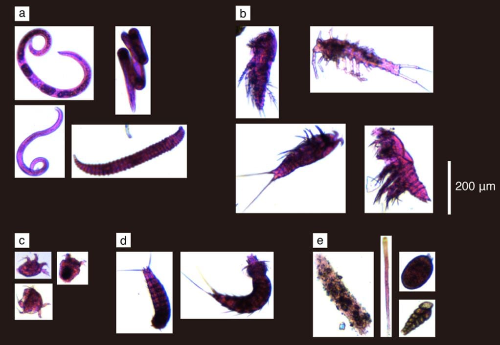
Pictured above: Meiobenthos imaged by the FlowCam. Organic matter was stained with Rose Bengal to easily differentiate meiobenthos from inorganic particulates, such as sediment. Imaged organisms are labeled as follows: a) Nematoda; b) Copepoda; c) Nauplius larvae; d) Kinorhyncha; e) Foraminifera. Credit: Kitahashi et al. (2018).
Meiobenthos are small, benthic invertebrates often used as indicators of anthropogenic influence and other natural disturbances. They play a primary role in sediment nutrient cycling and stability in benthic ecosystems.
Optical microscopy, which is labor-intensive and time-consuming, is often the primary technology utilized for analysis of meiobenthos. In this study, Kitahashi et al. developed a method to use the FlowCam and VisualSpreadsheet® for analysis of these small, benthic invertebrates.
Kitahashi et al. report strategies to prevent flow cell clogging, preserve live organisms through analysis, and discriminate organic matter from inorganic matter in dealing with sediment-rich liquid samples.
In the end, they found that the FlowCam:
" 1) Enabled sufficient meiobenthic images to be obtained to allow the identification and classification of specimens at high taxonomic levels.
2) Obtained comparable numbers of individuals to traditional methods.
3) Has the potential to rapidly process large volumes of meiobenthos samples that are required when monitoring seasonal and spatial variation in ocean ecosystems and conducting long-term environmental impact assessments. "
Citation:
Kitahashi, T., Watanabe, H.K., Tsuchiya, M., Yamamoto, H., Yamamota H., 2018, A new method for acquiring images of meiobenthic images using the FlowCam. MethodsX 5, 1330-1335 (2018).


