Ever wondered how a FlowCam works, or how it compares to a traditional microscope? Read further to learn about the basic mechanics that all of our instruments use.

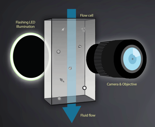
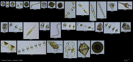
Flow imaging microscopy uses digital images to measure the size and shape of each particle. Essentially, the operator in classical microscopy is replaced by a computer to extract the information from the images. The liquid sample containing the particles streams through the flow cell past the microscope optics. Thousands of particle images are captured per second by the camera.
To capture sharp images of moving particles, they are “frozen” in space using a strobed illumination source combined synchronously with a very short shutter speed. As each frame of the camera’s field of view is captured, the software extracts the particle images from the background and stores
them.
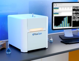
The intelligent paired software VisualSpreadsheet allows you to sort and filter the data based on the images captured by the camera. No longer constrained to just data, you can now see images of your particles and make informed decisions about what's in your sample, formulation, water or material.


