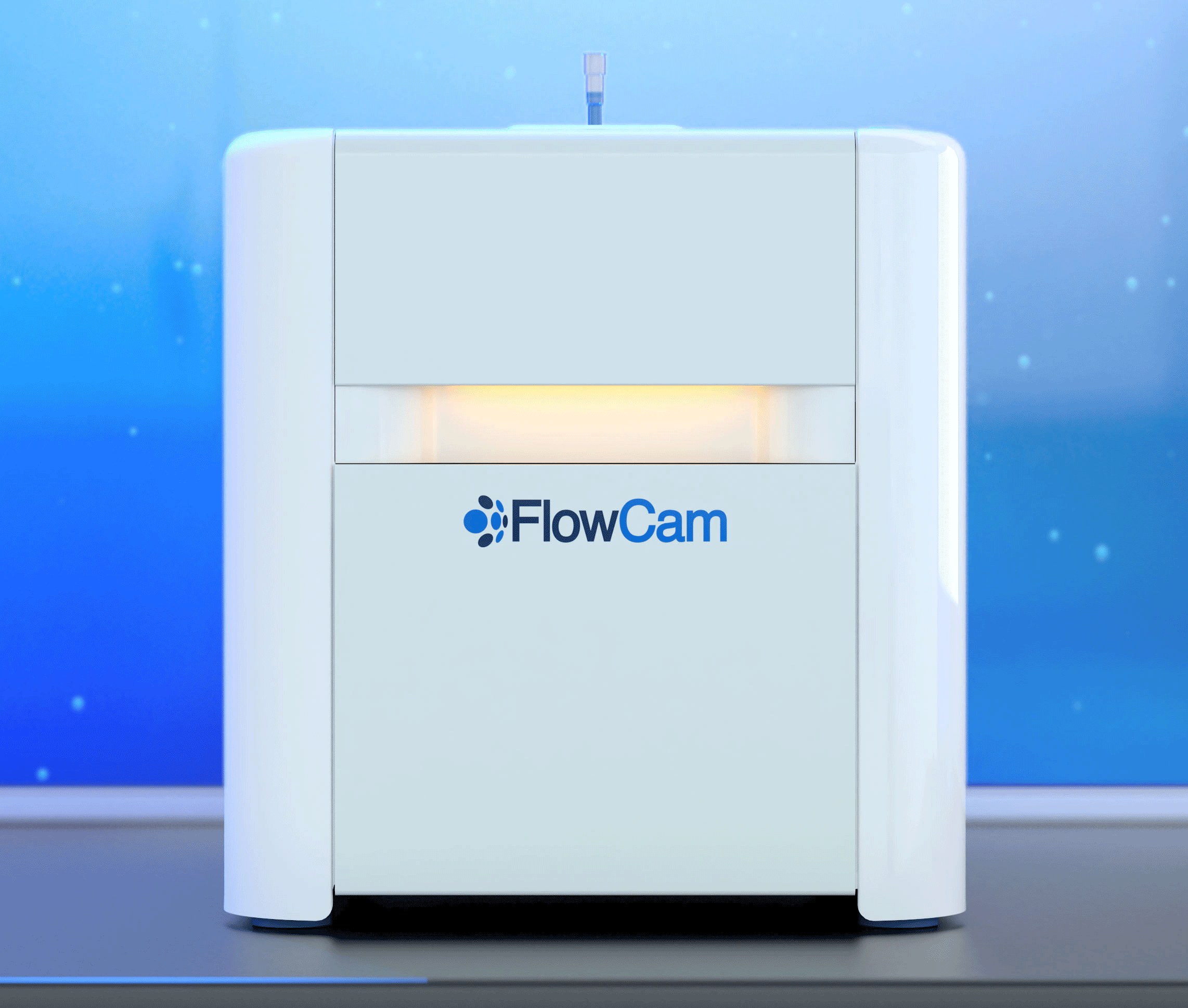
Have you ever wondered how FlowCam works, or how it compares to a traditional microscope?
FlowCam instruments use flow imaging microscopy (FIM) technology. FIM combines the benefits of traditional microscopy and high-throughput particle analyzers by capturing images of particles and microorganisms in a microfluidic channel under flow.
To capture sharp images of moving particles as they pass through a flow cell, they are “frozen” in space using a strobed illumination source combined synchronously with a very short shutter speed. As each frame of the camera’s field of view is captured, the software extracts the particle images from the background and stores them. Thousands of particle images are captured per second by the camera.

How it works:
-
A sample is manually or automatically loaded into the injection port
-
A high-precision syringe draws the sample into an optical flow cell
-
A fluidics sensor initiates data acquisition
-
A high-speed camera records images of the full width and depth of the flow cell as the sample flows through the optical field of view
-
Particle images are segmented from the camera images and captured in real-time as they flow through the flow cell
-
Data is further analyzed, grouped, and filtered post-acquisition
After a sample has been run, FlowCam's intelligent paired software, VisualSpreadsheet, essentially replaces the human operator in traditional microscopy by extracting over 40 parameters measured from the image data recorded by the camera. You are then able to use software filters to group, analyze, and visualize the collected data. No longer constrained to just count and size numbers provided by volumetric particle analysis techniques, with FlowCam you can see images of your particles to make informed decisions about what's in your sample.
Learn more about Flow Imaging Microscopy and how it compares to other particle analysis methods in our eBook:
And watch this video to see FlowCam in action:


