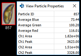
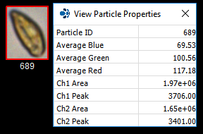
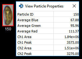
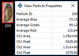
The FlowCam's "trigger-mode" enables the user to capture individual images of excited, fluorescing particles every time one passes the camera. Cyanobacteria and Cryptophyta contain the photopigments chlorophyll and phycobiliproteins. These photopigments can be excited using a FlowCam equipped with a 532 nm laser. When the FlowCam is set to "trigger-mode", each fluorescing particle that passes the camera view "triggers" the FlowCam to capture its image.
|
|
|
|
|
|
Pictured above: Cryptophyta images captured by the FlowCam using trigger mode. Relative fluorescence values for each cell denoted as Ch1 (chlorophyll) and Ch2 (phycobiliproteins) peaks. These relative fluorescence values are used to differentiate between phytoplankton with different fluorescent properties as captured by the FlowCam.
FIT team member Kay Johnson traveled to the University of Algarve in Faro, Portugal where she taught a group of aquatic researchers how to operate a FlowCam equipped with a 532 nm laser. During this training session, her group obtained a plankton tow in nearby coastal lagoon Ria Formosa and "FlowCammed it" using trigger mode. Both diatoms and Cryptophyta were imaged and relative fluorescent data was obtained for each type of phytoplankton.
The FlowCam with the green laser (532 nm) contains two photomultiplier tubes: CH1 with a 650LP filter and CH2 with a 575M30 filter. Chlorophyll triggers the FlowCam’s camera through the CH1 detector, while phycobiliproteins (phycoerythrin) triggers the camera through the CH2 detector. The FlowCam uses each image it captures to obtain quantitative measurements. This data is used by VisualSpreadsheet to evaluate trends and perform statistical analyses and calculations for each sample run. 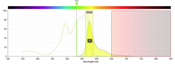 Pictured above: A FlowCam equipped with a green (532 nm) laser excites phycoerythrin (yellow curve) and chlorophyll (spectrum not shown). The FlowCam's CH2 PMT captures the peak phycoerythrin emissions (yellow shaded box) and CH1 PMT captures chlorophyll emissions (red shaded box) (Credit: Thermofisher)


