Ever wonder if you could use your FlowCam to study microplastics? The state of California thinks you can! In its 2021 Draft Microplastics in Drinking Water Policy Handbook, the state cites FlowCam as a viable surrogate for spectroscopic methods like FTIR and Raman Microscopy. Alongside these modalities, our particle imaging analysis of microplastics demonstrates the value FlowCam offers.
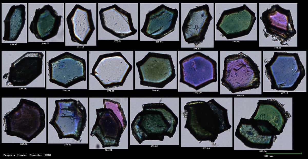
Pictured here: Multicolored plastic glitter, as imaged by FlowCam with 4X objective.
The Interstate Technology & Regulatory Council (ITRC) presented a training course that informed state regulators, environmental consultants, and community and tribal stakeholders of the pervasiveness of microplastics in the environment. Additionally, the ITRC and the Australasian Land and Groundwater Association (ALGA) held an expert panel on the latest microplastics policies, regulations, and research from the US, Australia, and other countries, further emphasizing the severity of the contaminant worldwide.
We know microplastics research is an important topic for many of you - and we're here to tell you that FlowCam can help! While microplastic speciation capabilities are being developed, FlowCam currently provides total microplastic concentration values in addition to these advantages:
- Rapid analysis: process 1 mL in 5 minutes
- Particle size distribution: image and count particles from 2 µm to 1 mm (depending on configuration)
- High reproducibility: accurately count and image all particles in a sample, including microplastics.
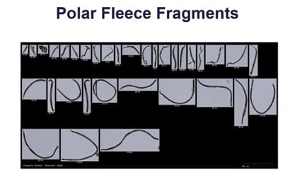
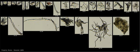
It can be challenging to identify microplastics compared to other particles. As shown in the image below, plastic polar fleece fragments appear like detritus in a water sample when imaged by FlowCam.
Photo: Woods et al., 2018
Download our new application note
Monitoring Microplastics Using FlowCam
Extra sample preparation steps, like the digestion and staining methods, might be needed to optimize your FlowCam analysis. We have collected the following resources for you to reference as you optimize your microplastic samples for FlowCam analysis. We also invite you to contact us if you would like to discuss method-development for your microplastics application.
- Two recently published research studies from Ewha Womans University in Seoul, Korea, are significant because they explore FlowCam's capabilities in analyzing polyethylene (PE) and polyamide (PA) microplastics.
FlowCam's Role in Microplastic's Analysis for Enhanced Environmental Solutions - A journal article published in 2019 by Lv et al. showed improved absorption of fluorescent dye to microplastic surfaces. For FlowCam users with a laser, consider using this method in conjunction with FlowCam analysis in trigger mode to detect fluorescing particles.
- Using a digestion method will remove organic material from your sample, leaving only inorganics. Refer to this method optimized for the digestion of wastewater samples for an example methodology (Cunsolo et al., 2021)
- A study by Woods et al. observed how the Mytilus edulis, or the blue mussel, interacts with microplastic fibers at concentrations representative of coastal waters (3 to 30 particles per mL). Microplastic fibers were identified and enumerated using the FlowCam imaging flow cytometer.
Microplastic Fiber uptake, ingestion, and egestion rates in the blue mussel (Mytilus edulis) - A study by Lorenz et al (2017) "Using the FlowCam to Validate an Enzymatic Digestion Protocol Applied to Assess the Occurrence of Microplastics in the Southern North Sea"
- From the Ocean Sciences Meeting in 2018, a poster presentation: Use of Imaging Flow Cytometry (FlowCam) in the Study of Microplastics.


