The recently launched FlowCam Nano extends well-established flow imaging microscopy technology for subvisible particle analysis into the submicron size range. With its unique ability to capture images and analyze particles from 300 nm to 2 µm in diameter, FlowCam Nano technology offers the ability to bridge the gap between different particle analysis techniques.
 FlowCam was originally invented at Bigelow Laboratory for Ocean Sciences (BLOS) in 1997 by Chris Sieracki, and at the time, it represented the world’s first imaging flow cytometer. It revolutionized the tedious and slow process of manual examination of phytoplankton via microscopy by providing a semi-automated method to rapidly count, measure, and analyze individual cells and particles in a fluid sample using digital images.
Pictured at right, inventor Chris Sieracki with a FlowCam prototype that was deployed on the dock at Bigelow Labs in the late 90s.
The groundbreaking paper “An imaging-in-flow system for automated analysis of marine microplankton” was published in 1998, setting the stage for the founding of Fluid Imaging Technologies in East Boothbay, Maine in 1999.
Over the next 20+ years, we expanded our FlowCam line of instruments to broaden the range of imaging capabilities and to expand the applications for flow imaging microscopy beyond oceanographic research. Today FlowCam applications range from biotherapeutic and cell research applications to harmful algal bloom monitoring in marine and freshwater ecosystems to materials characterization and more. Visit our applications page to see the full range of what is possible with FlowCam.
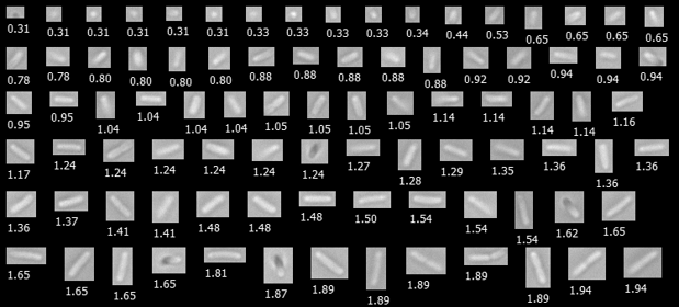
Pictured above: E. coli cells, some of the smallest organisms detectable by FlowCam Nano (diameter shown in µm)
While FlowCam Nano offers images and particle counts in the submicron range down to 300 nm, at the other end of the size spectrum, FlowCam Macro is used to image particles and organisms in the visible size range (up to 5mm), such as industrial microspheres, fibers, as well as zooplankton.
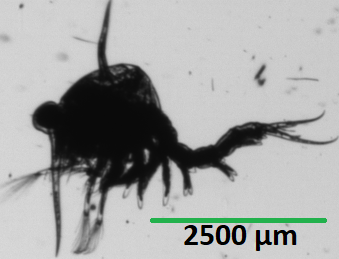
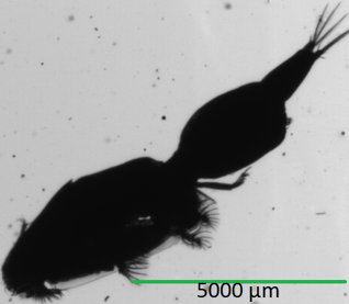
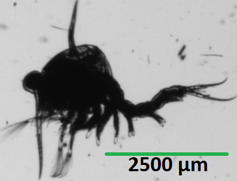
Pictured below: Zooplankton from the Gulf of Maine, some of the largest organisms detectable by FlowCam Macro. Various species of sea lice are found in the Gulf of Maine, causing problems with both the commercial and recreational fisheries, as well as in finfish aquaculture. Green Crabs are an invasive species in Maine that are now causing significant problems with the soft-shell clam industry, which represents Maine's third most valuable marine species. These images were captured by Bigelow Labs, where FlowCam was originally developed. Bigelow now uses several different FlowCam models to further their research.
 |
 |
| Sea Lice with an Area Based Diameter (ABD) of 2500µm |
Green crab larvae with an Area Based Diameter (ABD) of 1500µm |
Learn more about the various FlowCam models:
 |
 |
 |
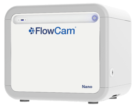
FlowCam NanoFor particles 300 nm to 2µm |
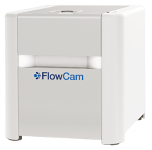
FlowCam 8000For particles 2 µm to 1 mm |
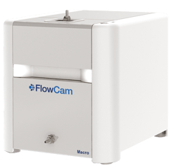
FlowCam MacroFor particles 300 µm to 5 mm |


