Situated on the picturesque Gulf Coast of Florida, Eckerd College boasts one of the nation's most esteemed undergraduate marine science programs. Among its faculty are Drs. Amy Suida and Shannon Gowans who recently joined forces to conduct a groundbreaking study on microplastics in Tampa Bay. Their research delves into the vital topic of microplastic ingestion by two distinct creatures - the magnificent Florida manatee and the minuscule copepod Acartia tonsa. We had the privilege of speaking with Amy and Shannon to gain insights into their undergraduate research programs and their experience with the innovative FlowCam technology.
How did you first learn about FlowCam?
We were collaborating with a thesis student on a research project to determine if copepods would avoid contaminated microplastics. One of the challenges we faced was accurately quantifying the similarly sized phytoplankton and plastic microbeads during the ingestion experiments. Fortunately, FlowCam proved the ideal solution, allowing us to observe the particles and differentiate between beads and diatoms. Additionally, we were able to monitor the aggregation of the beads throughout the experiments. The visualization capabilities of FlowCam were invaluable in revealing that the beads we used for the ingestion experiment were forming aggregates, which we could not detect with a Coulter Counter. This discovery became crucial as bead aggregation could potentially affect ingestion rates and contamination levels.
How are you currently using FlowCam?
 We recently incorporated a 2X objective into our FlowCam system to enhance our imaging capabilities (beyond 4X, 10X & 20X). This addition allows us to capture high-resolution images of phytoplankton and zooplankton, which we are thrilled to share with our students in the biological oceanography course. We are particularly excited about the opportunities presented for method development in analyzing environmental microplastics samples. Previously, our method involved staining the microplastics, using a microscope to capture images, and stitching those images together to quantify the plastic particles. This process was quite laborious. However, with FlowCam's advanced imaging capabilities, we can streamline and improve this process significantly. FlowCam delivers superior imaging quality, making it an invaluable tool for our research.
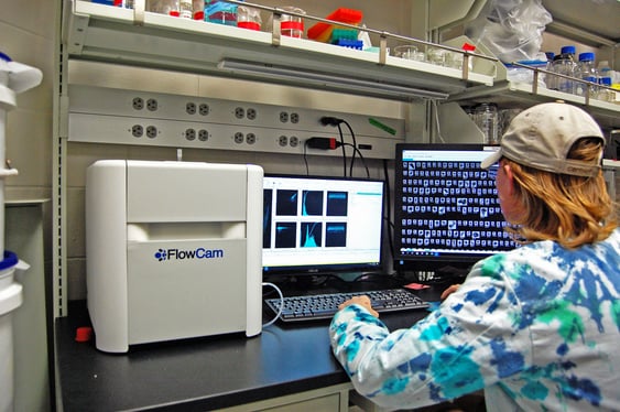
Pictured above: Eckerd College student uses FlowCam in their lab.
What challenges has FlowCam helped you solve?
The particle visualization capabilities of FlowCam are truly remarkable. Previously, the photos captured did not provide enough detail to analyze the particles accurately, so we are limited to obtaining information on particle quantity. However, with FlowCam, we can go beyond that and evaluate the shape and types of particles in much greater detail. By capturing higher-resolution images of each particle, FlowCam allows us to understand the microplastic particles in Tampa Bay.
How has FlowCam improved your workflow?
As we continue to refine our workflow, we are thrilled to provide our undergraduate students with the chance to learn about method development, an opportunity typically reserved for graduate students. In the past, we have had multiple undergraduate students utilize FlowCam for culture-based samples, and currently, one of our students is working on this project for environmental samples.
Interested in learning more about aquatic applications with FlowCam? Visit our Resource Library.


