“Is it automatically taking pictures of our sample?”
“Can we sort our data by size?”
“Will it help me find similar-looking images?”
With FlowCam, the answer to each of these questions is “Yes”.
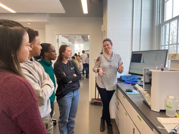
Typically, these are questions that we get asked by professional scientists and graduate-level researchers. But during our February visit to Bates College, it was undergraduate students asking all the questions.
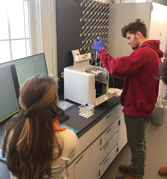
Bates, a leading liberal arts college in Lewiston, Maine, is known for its Biological and Biomedical Science programs and low student-teacher ratios. We had the privilege of visiting the school earlier this month in order to demonstrate the value of FlowCam as part of an undergraduate Biology curriculum.
After more than two years of virtual seminars during the pandemic, this was the first time since 2020 that we had the opportunity to bring FlowCam directly into an undergraduate lab, and we had so much fun doing it!
FlowCam Sample Analysis
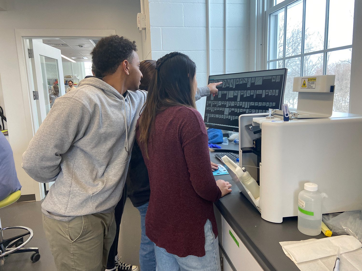
We collaborated with students and professors in two sections of a level 200 course, Molecules to Ecosystems. Each section included 12 groups of four students. After being presented with a brief overview, each student team ran a sample of rumen fluid through the FlowCam for analysis. Each group was asked to load 500 µL of a pre-filtered and diluted rumen fluid sample into the FlowCam instrument and then observe the FlowCam image and data acquisition process.
Microscopic analysis of rumen fluid is typically used to assess protozoan livelihood in a cow's digestive system. Traditional light microscopic analysis however is time-consuming and can only interrogate a very small sample of the enormous microbial content in the rumen. Using flow imaging microscopy, FlowCam allowed the students to immediately visualize the microbial diversity of the rumen fluid. FlowCam's rapid imaging, sizing, and counting of protozoa and other organisms also helped the students gain a firsthand appreciation for how the rumen ecosystem might function to support cow health.
Comparing Technologies
After the samples were run, students were asked to select images of microbes and other rumen fluid particles that they found most interesting to build FlowCam collages. FlowCam data on the physical properties of their selected images were exported as CSV files and made available to the students to analyze.

Pictured above: a collage of rumen fluid particles built by a Bates student
They were then asked to answer the following questions:
"How did FlowCam help you understand or appreciate rumen fluid as an ecosystem?"
"What did the FlowCam allow you to see that your microscopy images didn’t?"
and "What did you see under the microscope that you didn’t see with the FlowCam?"
Here are some of the students' responses:
"In microscopy photos, we can observe the partial view at one time, but we cannot get a holistic view of the whole ecosystem (rumen)."
"It was very interesting getting to work firsthand with a FlowCam. I found it fascinating to see how quickly the FlowCam could pick up so many images of the different bacteria, protozoa, and food pieces all found in the rumen. It really helped demonstrate just how much there is living in the rumen microbiome. We only placed a small drop of rumen sample on the FlowCam but the camera produced hundreds of images of bacteria, protozoa, and other things in the rumen."
"The FlowCam images made it easy to see the diversity of microorganisms present in the rumen fluid. This is an advantage over the microscopy images because under a microscope you’re limited to seeing what you can find by panning around the slide and any given image can only include so much. FlowCam also makes it easier to compare the size of structures."
"FlowCam's high-resolution imaging capabilities can pick up on minute changes between various cell types, aiding in the more precise identification and classification of microbes...Traditional microscopy, on the other hand, gave us the ability to see the three-dimensional organization of cells and tissues, giving us more precise information on their interior structure and shape."
Along with the course professors, we greatly enjoyed observing the students' enthusiasm for hands-on experience with a new technology. FlowCam gave students a fun way to see for themselves that the cow's rumen is indeed an ecosystem rich in diverse microorganisms. FlowCam’s ease of use makes it an accessible teaching and learning instrument with great value for engaging students in new concepts and for supporting authentic research experiences in undergraduate labs.
Bates College faculty are thinking of ways to use FlowCam for student research on soil and river ecology. As part of place-based research studies, students would take water and soil samples from a local river to assess the effects of an upcoming dam removal and bank restoration project.


