FlowCam applications are wide-ranging, from aquatic systems monitoring and marine organism research to cell therapy development and advanced materials characterization. While particles in samples from these diverse areas are distinct, FlowCam provides the highest quality image-based analysis when the optimal instrument conditions are used.
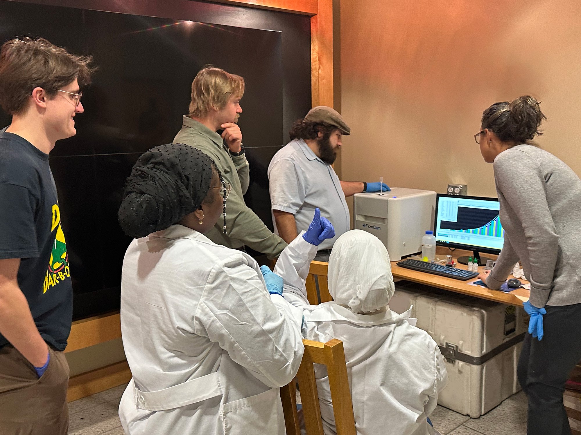
 Recently, we had the fortunate opportunity to explore FlowCam’s versatility across several application areas all in one visit to the MDI Biological Laboratory (MDIBL) in Bar Harbor, Maine. MDI Bio Lab is a non-profit, international hub for the science of aging and regeneration. Its state-of-the-art facilities support 10 research groups whose faculty and students are pushing the frontiers of biomedical science, discovering new pathways to health and longevity.
In a single afternoon, we analyzed water samples from the Animal Resources Core, kidney organoids from the research lab of institute president Hermann Haller, M.D., and zebrafish embryos from the Kathryn W. Davis Center for Regenerative Biology and Aging. With so many different types of “particles” to look at in one afternoon, getting started with the most appropriate FlowCam instrument and accessory pairings was key!
Achieving success requires a keen understanding of the capabilities of the new instrumentation and operating methods that are appropriately tuned for your specific application. Hardware configuration, software settings customization, and sample preparation are essential considerations when getting started.
Aquaculture Water Samples
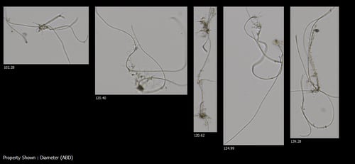 Using the FlowCam 8000, with a field of view (FOV) 600 flow cell, and a magnifying objective lens of 4X, we analyzed water samples taken before and after new filters were installed in the aquaculture tank systems. The goal of this analysis was to assess water quality improvement in response to filter changes. Therefore, the samples were directly analyzed by FlowCam 8000 after collection without any special sample preparation. It was interesting to notice a few fiber-like particles in samples collected from tank locations near the filter outlet. This observation might inform filter preparation methods and water sample collection protocols.
Organoid Tissue Cultures
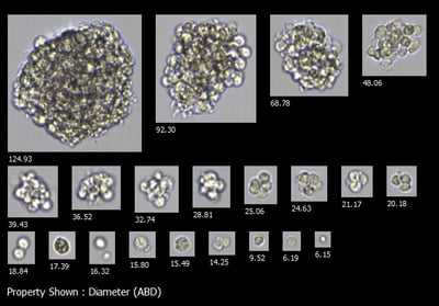 Using FlowCam 8000 again but with different accessory pairings, this time with the FOV 100 and 10X magnification lens, we helped students from Colby College assess the effects of the different protocols they used to dissociate cells from early organoid tissue cultures.
Zebrafish Embryos
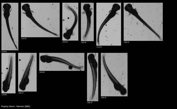
With FlowCam Macro, we were able to capture full-body zebrafish embryo images! With a broader particle size range of up to 5mm and lower magnification specifications, FlowCam Macro was the instrument of choice to attain high-resolution images showing intricate features of the developing zebrafish embryos. Below is a collage file of Zebrafish embryo images captured at 0.5X on FlowCam Macro.
Having the opportunity to implement state-of-the-art technology to improve efficiency and strengthen the value of your analytical work is invigorating, knowing you will now be able to deliver on your goals and objectives.
For more information on accessory pairings and application-specific context settings, see the following technical note: FlowCam 8000 Series Configuration Guide.
For information about FlowCam instrument particle size range and magnification specifications, visit the FlowCam Macro product page.


